Diabetes eye exams
Description
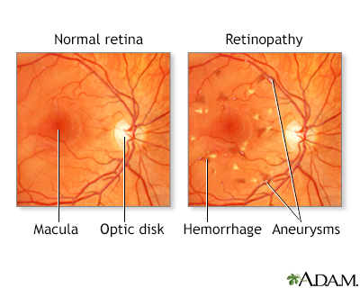
Diabetes can harm your eyes. It can damage the small blood vessels in your retina, the back wall of your eyeball. This condition is called diabetic retinopathy.
Diabetes also increases your risk of glaucoma and other eye problems.
You may not notice that your eyes are damaged until the problem is very bad. Your eye doctor can catch problems early if you get regular eye exams. This is very important. The early stages of diabetic retinopathy don't cause changes in vision and you won't have symptoms. Only an eye exam can detect the problem, so that steps can be taken to prevent the eye damage from getting worse.
Patient Education Video: Diabetes
Alternative Names
Diabetic retinopathy - eye exams; Diabetes - eye exams; Glaucoma - diabetic eye exam; Macular edema - diabetic eye exam
You Need Regular Eye Exams
Even if the health care provider who takes care of your diabetes checks your eyes, you need an eye exam every 1 to 2 years by an eye doctor who takes care of people with diabetes. An eye doctor has equipment that can check the back of your eye much better than your regular provider can.
If you have eye problems because of diabetes, you will probably see your eye doctor more often. You may need special treatment to prevent your eye problems from getting worse.
You may see two different types of eye doctors:
- An ophthalmologist is a medical doctor who is an eye specialist.
- An optometrist is a doctor of optometry. If you develop eye disease caused by diabetes, you will likely also see an ophthalmologist.
What is a Dilated Retinal Exam?
The doctor will check your vision using a chart of random letters of different sizes. This is called the Snellen chart.
You will then be given eye drops to widen (dilate) the pupils of your eyes so that the doctor can better see the back of the eye. You may feel stinging when the drops are first placed. You may have a metallic taste in your mouth.
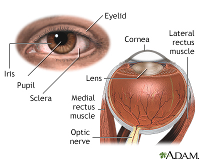
To see the back of your eye, the doctor looks through a special magnifying glass using a bright light. The doctor can then see areas that may be damaged by diabetes:
- Blood vessels in the front or middle parts of the eye
- The back of the eye
- The optic nerve area
Another device called a slit lamp is used to see the clear surface of the eye (cornea).
The doctor may take photos of the back of your eye to get a more detailed exam. This exam is called a digital retinal scan (or imaging). A special camera is used to take photos of your retina without dilating your eyes. The doctor then views the photos and lets you know if you need more tests or treatment.
After Your eye Exam
If you had drops to dilate your eyes, your vision will be blurred for about 6 hours. It will be harder to focus on things that are near. You should have someone drive you home.
Also, sunlight can damage your eye more easily when your pupils are dilated. Wear dark glasses or shade your eyes until the effects of the drops wear off.
Gallery
References
American Academy of Ophthalmology website. Diabetic retinopathy PPP 2019. www.aao.org/preferred-practice-pattern/diabetic-retinopathy-ppp. Updated October 2019. Accessed October 31, 2022.
American Diabetes Association website. 12. Retinopathy, neuropathy, and foot care -2022. Diabetes Care. 2022;45(Suppl 1):S185-S194. PMID: 34964887 pubmed.ncbi.nlm.nih.gov/34964887/.
Brownlee M, Aiello LP, Sun JK, et al. Complications of diabetes mellitus. In: Melmed S, Auchus, RJ, Goldfine AB, Koenig RJ, Rosen CJ, eds. Williams Textbook of Endocrinology. 14th ed. Philadelphia, PA: Elsevier; 2020:chap 37.
Skugor M. Diabetes mellitus. In: Sadda SVR, Saraff D, Freund KB, et al, eds. Ryan's Retina. 7th ed. Philadelphia, PA: Elsevier; 2023:chap 48.