"Thanks to Dr. Friedman, I can have a conversation with my husband without being in pain, read books to my baby girl, Amber, without flinching, and I just love to get their kisses." - Wanda, Crawfordville, Florida

Update your location to show providers, locations, and services closest to you.

At University of Florida Health, we perform more than 100 trigeminal neuralgia procedures each year. Surgery for the treatment of trigeminal neuralgia is performed by William Friedman, MD.
Trigeminal neuralgia is commonly referred to as “tic douloureux” or just “tic." When severe, it's the most excruciating pain known to man. This nerve pain most frequently involves the lower lip and lower teeth or the upper lip and cheek,. But it also may involve the nose and the area above the eye.
Routine pain medications often are ineffective in controlling this neuropathic pain. The most effective drug for controlling this neurological disorder is Tegretol. However, it often caused side effects such as drowsiness and sleepiness, mental slowness or imbalance. More serious side effects, such as blood and liver problems, have also been reported.
Dilantin, Baclofen and Neurontin are also used for trigeminal neuralgia but are generally less effective. Almost all patients with trigeminal neuralgia pain initially obtain excellent relief from Tegretol, so we routinely recommend it. However, most patients find that, over time, larger and larger doses of medication are required to stop pain recurrence, and that it eventually becomes ineffective or causes intolerable side effects. Fortunately, here at UF Health, we have several excellent surgical procedures to offer patients.
In planning a surgical procedure for trigeminal neuralgia, it's important to determine which branch of the trigeminal nerve is involved. The trigeminal nerve has three branches which correspond to the upper, middle and lower portions of the face.
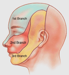
The first (upper) branch includes the eye, eyebrow and forehead. The second (middle) branch corresponds to the upper lip, upper teeth, upper gum, cheek, lower eyelid and side of the nose. The third (lower) branch involves the lower lip, lower teeth, lower gum and one side of the tongue. It also includes a narrow area that extends from the lower jaw in front of the ear to the side of the head.
Trigeminal neuralgia most commonly involves the middle (second) and lower (third) branches, but may involve the upper (first) branch alone, any two branches or all three branches.
After identifying the affected branch, the next decision is the selection of a surgical procedure. The most common operations we perform for trigeminal neuralgia are vascular decompression, percutaneous stereotactic radiofrequency lesion and radiosurgery procedures. Our UF Health experts are pioneers in radiosurgery for many other diseases and are proud to offer this operation for patients experiencing trigeminal neuralgia pain.
The vascular decompression procedure is a major surgery designed to minimize facial numbness as part of the treatment. The radiofrequency procedure is a more minor, needle-type procedure. It's designed to relieve pain by causing facial numbness in in the region of the branch or branches involved in the nerve pain.
Injection therapy to offer temporary pain relief also is available. Novocaine or Xylocaine injections, such as those used in dentistry, can numb the painful area for a few hours. Alcohol and glycerol injections and other means of destroying part of the trigeminal nerve also may be considered.
With almost every type of injection and surgical procedure, including the radiofrequency lesion procedure, there is a small chance the pain will recur. If the facial numbness wears off and the pain returns, the procedure can be repeated.
The vascular decompression procedure is the surgery recommended for a healthy person who does not want facial numbness and is willing to accept a major surgery through the skull. It relieves trigeminal neuralgia by placing a small pad between the trigeminal nerve and the blood vessels next to the nerve.
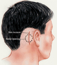
The vascular decompression surgery requires making an incision in the back of the head, creating a small hole in the skull and lifting an edge of the brain to expose the trigeminal nerve which is located approximately two inches deep. The incision is made behind the ear on the side of the head where the patient feels pain.
The blood vessels that press on the nerve fibers when the nerve leaves the brain are exposed and pushed away from the nerve. A small pad is inserted between the nerve and the blood vessels. This relieves the intense pain in most patients.
The surgery requires a general anesthetic and involves a small risk of complications. These risks include facial numbness, facial weakness, hearing loss, double vision, infection, bleeding, stroke, hydrocephalus, CSF leak, and other neurological issues.
Recurrent facial pain following the surgery occurs in about 30 percent of patients. If the pain recurs another vascular decompression surgery or a radiofrequency lesion procedure (described later) may be required.
The preoperative evaluation is done by the doctor and the anesthesia service in the clinic as an outpatient prior to the vascular decompression surgery. Prior to the surgery, the patient will be admitted to the hospital on the day of your surgery. The procedure typically takes about 45 minutes. Most patients spend two nights in the hospital. Intensive care is usually not necessary. Patients should plan on taking approximately two weeks off work after leaving the hospital and expect a follow-up appointment within four weeks after leaving the hospital.
The other procedure we commonly perform to treat trigeminal neuralgia is called percutaneous stereotactic radiofrequency destruction of the trigeminal nerve. The term “percutaneous” means the treatment is performed with a needle passed through the skin. The term “stereotactic” refers to the fact the needle is directed by X-ray control. The term “radiofrequency” refers to the radiofrequency heating current which is used to destroy the nerve.
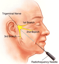
Relief of the trigeminal neuralgia by this method involves making the region of the pain permanently numb. The RFL procedure is performed in the operating room with the patient lying horizontally on their back. A needle is passed, under X-ray control, into the cheek on the side of the face where the patient feels nerve pain and through a small, natural opening in the base of the skull into the trigeminal nerve.
The patient is placed under general anesthesia for a few minutes during the insertion of the needle and during other portions of the surgery. This is accomplished with a medication similar to Pentothal called Brevital, which results in a very brief period of sleep.
After inserting the needle, the patient is awakened and a small electric current is passed through the needle causing tingling in the face. When the needle is positioned so the tingling occurs in the area of the tic pain, the patient is put to sleep again and a radiofrequency current is passed through the needle to destroy part of the nerve.
The patient is awakened a few minutes after completing the nerve lesion and is checked to determine if there is enough numbness in the face to give pain relief. The radiofrequency lesion procedure is repeated with the patient asleep until it has resulted in the desired facial numbness.
In most cases, the X-ray portion of the procedure takes approximately 15 minutes. When the procedure is completed, the patient goes to the recovery room for about two hours after which they can go home. They are usually able to eat the next meal. The facial numbness with this procedure usually is permanent. Should the facial numbness wear off, there is a chance of recurrent tic pain in which case the procedure can be repeated.
Several side effects may follow the procedure. The effects described below apply to the radio frequency lesion procedure. Please consult with your health care provider for additional details about what to expect with your individual care. Following the radio frequency lesion procedure, you may experience numbness or have an unpleasant or painful sensation similar to the way it feels after a Novocain injection.
It's possible to stick a pin into the numbed area without the person feeling it, yet the patient may describe this sensation with words such as it “tingles,” “burns,” “draws,” “pulls,” “crawls,” or it is “woody” or “stiff like cement.”
Some patients find the numb area seems irritated or aches. One or two percent of the patients will find this sensation more disagreeable than their original tic pain. However, an overwhelming majority feel the facial numbness is far better than the intense tic pain.
The second most common side effect is a weakness of the chewing muscles on the side of the head where the patient feels the pain. Many people describe the weakness as a change in their bite or as an inability to chew as hard on the side of the lesion. This weakness usually recovers six to eight months after the procedure.
The third side effect in an unwanted spread of the numbness to the adjacent branches of the nerve and to the eye. In some cases, we actually are trying to numb the area in and surrounding the eye because the pain is situated there. Numbness of the eye itself is not harmful, but if foreign matter enters the eye, the patient would not feel it. Inflammation, scarring of the cornea, and reduction or loss of vision could result. To prevent this, we recommend each patient inspects the eye regularly with a mirror and to see an eye doctor if the eye becomes red or appears irritated, even though he or she may not experience pain.
A few cases of double vision have been noted after the procedure, but none of these have been permanent. Rarely, meningitis or other neurological disorders may occur.
Patients are evaluated in the clinic the day before the procedure. Patients are instructed to go to the outpatient surgery desk on the morning of the surgery and are taken to the operating room from there. After the procedure, the patient remains in the recovery room for about two hours before being discharged to go home. The patient can return to work a day or two following the procedure.
To schedule an appointment to speak to our knowledgeable expert on this condition, please call (352) 273-6990 or complete the appointment consultation form.
"Thanks to Dr. Friedman, I can have a conversation with my husband without being in pain, read books to my baby girl, Amber, without flinching, and I just love to get their kisses." - Wanda, Crawfordville, Florida
In this video, patients describe their symptoms and outcomes and Dr. William Friedman describes diagnosis and treatment options for trigeminal neuralgia.


More than 900,000 adults in the United States are living with multiple sclerosis, according to the National Multiple Sclerosis Society. The multiple sclerosis…

Trigeminal neuralgia is a challenging condition that can cause excruciating pain in the face. The condition affects the trigeminal nerve, which is responsible…
