Chest CT
Definition
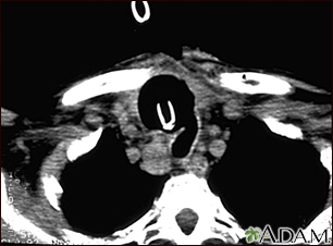
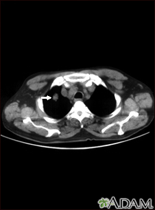
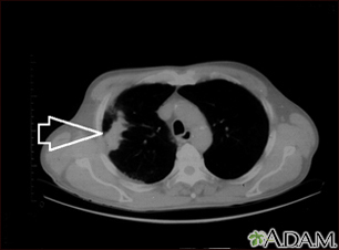
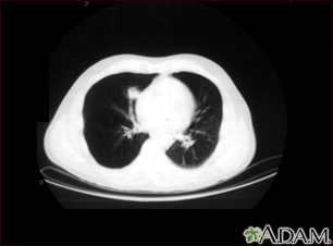
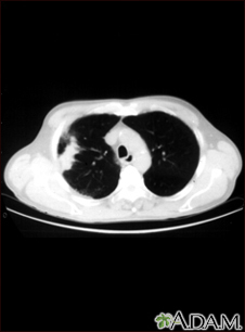
A chest CT (computed tomography) scan is an imaging method that uses x-rays to create cross-sectional pictures of the chest and upper abdomen.
Alternative Names
Thoracic CT; CT scan - lungs; CT scan - chest
How the Test is Performed
The test is done in the following way:
- You'll likely be asked to change into a hospital gown.
- You lie on a narrow table that slides into the center of the scanner. Once you are inside the scanner, the machine's x-ray beam rotates around you.
- You must be still during the exam, because movement causes blurred images. You may be told to hold your breath for short period of time.
The complete scan takes 30 seconds to a few minutes.
Certain CT scans require a special dye, called contrast, to be delivered into the body before the test starts. Contrast highlights specific areas inside the body and creates a clearer image. If your provider requests a CT scan with intravenous contrast, you will be given it through a vein (IV) in your arm or hand. A blood test to measure your kidney function may be done before the test. This test is to make sure your kidneys are healthy enough to filter the contrast.
You may be given medicine to help you relax during the test.
How to Prepare for the Test
Some people have allergies to IV contrast and may need to take medicine before their test to safely receive this substance.
If contrast is used, you may also be asked not to eat or drink anything for 4 to 6 hours before the test.
If you weigh more than 300 pounds (135 kilograms), have your health care provider contact the scanner operator before the exam. CT scanners have an upper weight limit of 450 pounds (204 kilograms). Some newer scanners can accommodate up to 680 pounds (308 kilograms). Because it is hard for x-rays to pass through metal, you will be asked to remove jewelry.
How the Test will Feel
Some people may have discomfort from lying on the hard table.
Contrast given through an IV may cause a slight burning sensation, a metallic taste in the mouth, and a warm flushing of the body. These sensations are normal and usually go away within a few seconds.
There is no recovery time, unless you were given medicine to relax. After a CT scan, you can go back to your normal diet, activity, and medicines.
Why the Test is Performed
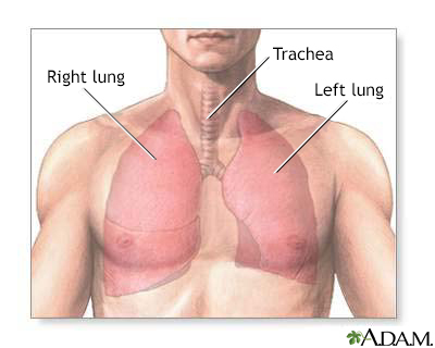
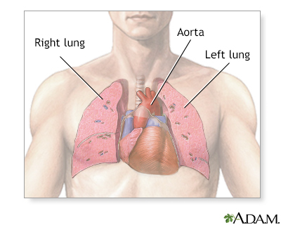
CT quickly creates detailed pictures of the body. The test may be used to get a better view of the structures inside the chest. A CT scan is one of the best ways of looking at soft tissues such as the heart and lungs.
A chest CT may be done:
- After a chest injury
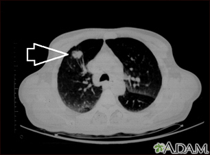
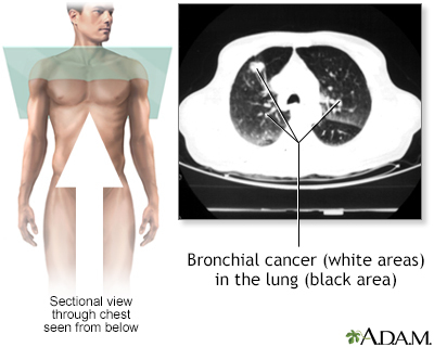
- When a tumor or mass (clump of cells) is suspected, including a solitary pulmonary nodule seen on a chest x-ray
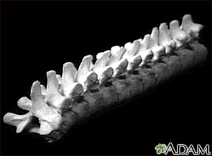
- To determine the size, shape, and position of organs in the chest and upper abdomen
- To look for bleeding or fluid collections in the lungs or other areas
- To look for infection or inflammation in the chest
- To look for blood clots in the lungs
- To look for scarring in the lungs
- To look for emphysema
What Abnormal Results Mean
Thoracic CT may show many disorders of the heart, lungs, mediastinum, or chest area, including:
- A tear in the wall, an abnormal widening or ballooning, or narrowing of the major artery carrying blood out of the heart (aorta)
- Other abnormal changes of the major blood vessels in the lungs or chest such as blood clots (pulmonary embolism)
- Buildup of blood or fluid around the heart
- Lung cancer or cancer that has spread to the lungs from elsewhere in the body
- Collection of fluid around the lungs (pleural effusion)
- Damage to, and widening of the large airways of the lungs (bronchiectasis)
- Enlarged lymph nodes
- Lung disorders in which lung tissues become inflamed and then damaged (intestinal lung disease)
- Pneumonia
- Esophageal cancer
- Lymphoma in the chest
- Tumors, nodules, or cysts in the chest
Risks
CT scans and other x-rays are strictly monitored and controlled to make sure they use the least amount of radiation. CT scans use low levels of ionizing radiation, which has the potential to cause cancer and other defects. However, the risk from any one scan is small. The risk increases as many more studies are done.
The most common type of contrast given into a vein contains iodine. If a person with an iodine allergy is given this type of contrast, nausea, sneezing, vomiting, itching, or hives may occur. In rare cases, the dye can cause a life-threatening allergic response called anaphylaxis. If you have any trouble breathing during the test, you should notify the scanner operator immediately. Scanners come with an intercom and speakers, so the operator can hear you at all times.
In people with kidney problems, the dye may have harmful effects on the kidneys. In these situations, special steps may be taken to make the contrast dye safer to use.
In spite of its risks, a CT scan may still be done if the benefits greatly outweigh the risks. For example, it can be more risky to not have the exam if your provider thinks you might have cancer.
Considerations
Gallery

References
Nair A, Barnett JL, Semple TR. Current status of thoracic imaging. In: Adam A, Dixon AK, Gillard JH, Schaefer-Prokop CM, eds. Grainger & Allison's Diagnostic Radiology. 7th ed. Philadelphia, PA: Elsevier; 2021:chap 1.
Shaqdan KW, Otrakji A, Sahani D. Safe use of contrast media. In: Abujudeh HH, Bruno MA, eds. Radiology Noninterpretive Skills: The Requisites. Philadelphia, PA: Elsevier; 2018:chap 20.