Skull x-ray
Definition
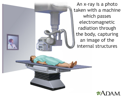
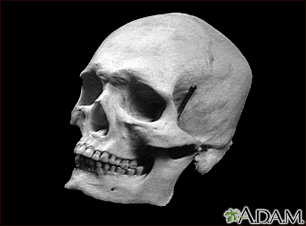
A skull x-ray is a picture of the bones surrounding the brain, including the facial bones, the nose, and the sinuses.
Alternative Names
X-ray - head; X-ray - skull; Skull radiography; Head x-ray
How the Test is Performed
You lie on the x-ray table or sit in a chair. Your head may be placed in different positions.
How to Prepare for the Test
Tell the health care provider if you are pregnant or think you are pregnant. Remove all jewelry.
How the Test will Feel
There is little or no discomfort during an x-ray. If there is a head injury, positioning the head may be uncomfortable.
Why the Test is Performed
Your doctor may order this x-ray if you have injured your skull. You may also have this x-ray if you have symptoms or signs of a structural problem inside the skull, such as a tumor or bleeding.
A skull x-ray is also used to evaluate an unusually shaped child's head.
Other conditions for which the test may be performed include:
- Teeth are not aligned properly (malocclusion of teeth)
- Infection of the mastoid bone (mastoiditis)
- Occupational hearing loss
- Middle ear infection (otitis media)
- Abnormal bone growth in the middle ear that causes hearing loss (otosclerosis)
- Pituitary tumor
- Sinus infection (sinusitis)
Sometimes skull x-rays are used to screen for foreign bodies that may interfere with other tests, such as an MRI scan.
A CT scan of the head is usually preferred to a skull x-ray to evaluate most head injuries or brain disorders. Skull x-rays are rarely used as the main test to diagnose such conditions.
What Abnormal Results Mean
Abnormal results may be due to:
- Fracture
- Tumor
- Breakdown (erosion) or calcium loss of the bone
- Movement of the soft tissues inside the skull
A skull x-ray may detect increased intracranial pressure and unusual skull structures that are present at birth (congenital).
Risks
There is low radiation exposure. X-rays are monitored and regulated to provide the minimum amount of radiation exposure needed to produce the image. Most experts feel that the risk is low compared with the benefits. Pregnant women and children are more sensitive to the risks associated with x-rays.
Gallery
References
Chernecky CC, Berger BJ. Radiography of skull, chest, and cervical spine - diagnostic. In: Chernecky CC, Berger BJ, eds. Laboratory Tests and Diagnostic Procedures. 6th ed. St Louis, MO: Elsevier Saunders; 2013:953-954.
Magee DJ, Manske RC. Head and face. In: Magee DJ, ed. Orthopedic Physical Assessment. 7th ed. Philadelphia, PA: Elsevier; 2021:chap 2.
Mettler FA Jr. Head and soft tissues of face and neck. In: Mettler FA, ed. Essentials of Radiology. 4th ed. Philadelphia, PA: Elsevier; 2019:chap 2.