Ophthalmoscopy
Definition
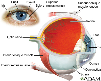
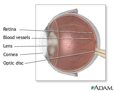
Ophthalmoscopy is an examination of the back part of the eye (fundus), which includes the retina, optic disc, choroid, and blood vessels.
Alternative Names
Funduscopy; Funduscopic exam
How the Test is Performed
There are different types of ophthalmoscopy.
- Direct ophthalmoscopy. You will be seated in a darkened room. The health care provider performs this exam by shining a beam of light through the pupil using an instrument called an ophthalmoscope. An ophthalmoscope is about the size of a flashlight. It has a light and different tiny lenses that allow the provider to view the back of the eyeball.
- Indirect ophthalmoscopy. You will either lie or sit in a semi-reclined position. The provider holds your eye open while shining a very bright light into the eye using an instrument worn on the head. (The instrument looks like a miner's light.) The provider views the back of the eye through a lens held close to your eye. Some pressure may be applied to the eye using a small, blunt probe. You will be asked to look in various directions. This exam is usually used to look for a detached retina.
- Slit-lamp ophthalmoscopy. You will sit in a chair with the instrument placed in front of you. You will be asked to rest your chin and forehead on a support to keep your head steady. The provider will use the microscope part of the slit lamp and a tiny lens placed close to the front of the eye. The provider can see about the same with this technique as with indirect ophthalmoscopy, but with higher magnification.
The ophthalmoscopy examination takes about 5 to 10 minutes.
How to Prepare for the Test
Indirect ophthalmoscopy and slit-lamp ophthalmoscopy are often performed after eyedrops are placed to widen (dilate) the pupils. Direct ophthalmoscopy and slit-lamp ophthalmoscopy can be performed with or without the pupil dilated.
You should tell your provider if you:
- Are allergic to any medicines
- Are taking any medicines
- Have glaucoma or a family history of glaucoma
How the Test will Feel
The bright light will be uncomfortable, but the test is not painful.
You may briefly see images after the light shines in your eyes. The light is brighter with indirect ophthalmoscopy, so the sensation of seeing after-images may be greater.
Pressure on the eye during indirect ophthalmoscopy may be slightly uncomfortable, but it should not be painful.
If eyedrops are used, they may sting briefly when placed in the eyes. You may also have an unusual taste in your mouth.
Why the Test is Performed
Ophthalmoscopy is done as part of a routine physical or complete eye examination.
It is used to detect and evaluate symptoms of retinal detachment or eye diseases such as glaucoma.
Ophthalmoscopy may also be done if you have signs or symptoms of high blood pressure, diabetes, or other diseases that affect the blood vessels.
Normal Results
The retina, blood vessels, and the optic disc appear normal.
What Abnormal Results Mean
Abnormal results may be seen on ophthalmoscopy with any of the following conditions:
- Viral inflammation of the retina (CMV retinitis)
- Diabetes
- Glaucoma
- High blood pressure
- Loss of sharp, central vision due to age-related macular degeneration
- Melanoma of the eye
- Optic nerve problems
- Retinal detachment (separation of the retina in the back of the eye from its supporting layers)
Ophthalmoscopy is considered to be 90% to 95% accurate. It can detect the early stages and effects of many serious diseases. For conditions that cannot be detected by ophthalmoscopy, there are other techniques and devices that may be helpful.
Risks
If you receive drops to dilate your eyes for the ophthalmoscopy, your vision will be blurred.
- Wear sunglasses to protect your eyes from sunlight, which can damage your eyes.
- Have someone drive you home.
- The drops usually wear off in several hours.
The test itself involves no risk. In rare cases, the dilating eyedrops cause:
- An attack of narrow-angle glaucoma
- Dizziness
- Dryness of the mouth
- Flushing
- Nausea and vomiting
If narrow-angle glaucoma is suspected, dilating drops are usually not used.
Gallery
References
Atebara NH, Miller D, Thall EH. Ophthalmic instruments. In: Yanoff M, Duker JS, eds. Ophthalmology. 5th ed. Philadelphia, PA: Elsevier; 2019:chap 2.5.
Ball JW, Dains JE, Flynn JA, Solomon BS, Stewart RW. Eyes. In: Ball JW, Dains JE, Flynn JA, Solomon BS, Stewart RW, eds. Seidel's Guide to Physical Examination. 9th ed. St Louis, MO: Elsevier; 2019:chap 12.
Chuck RS, Dunn SP, Flaxel CJ; American Academy of Ophthalmology Preferred Practice Pattern Committee, et al. Comprehensive adult medical eye evaluation preferred practice pattern. Ophthalmology. 2021;128(1):1-29. www.aaojournal.org/article/S0161-6420(20)31026-5/fulltext. Published November 12, 2020. Accessed March 2, 2021.
