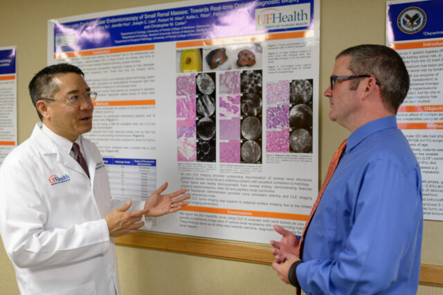
UF Health research shows that a tiny laser could help diagnose cancerous kidney tumors

University of Florida Health researchers are working to determine whether a tiny laser imaging probe can help detect whether a kidney tumor is cancerous or benign prior to subjecting a patient to an invasive needle biopsy or surgery.
When physicians find small tumors in the kidney, physicians currently perform a diagnostic needle biopsy or surgically remove the tumor to determine whether the tumor is cancerous or benign. Current imaging tests such as a CT, MRI or PET, or positron emission tomography, scan cannot precisely differentiate between the two. However, close to 20 percent of tumors that are surgically removed end up being benign, and as such, these patients may be undergoing unnecessary invasive procedures.
“A diagnostic needle biopsy that invades the tumor risks bleeding or leaching of cancer cells into surrounding tissues, or results in insufficient tissue to make an accurate diagnosis,” said Li-Ming Su, M.D., the David A. Cofrin professor and chair of the UF College of Medicine’s department of urology and the paper’s lead author. “We investigated whether a contact-based fiber-optic laser technology could be used to provide the same information as traditional invasive needle biopsies.”
Their results were published in February in the Journal of Urology.
The UF researchers are working with the company Mauna Kea Technologies to study the laser imaging technology. The technology, called confocal laser endomicroscopy, is basically a tiny microscope at the end of a 2.6-millimeter fiber-optic probe that uses a laser light to obtain real-time images of kidney tissues. The tissue must be bathed in fluorescent dye to illuminate cell structure and architecture when imaged by the laser light, said Su. A photo detector picks up reflected fluorescent light from the kidney tissues, and then produces a high-resolution image of the cellular architecture of the tissues being studied.
For this study, Su worked with researcher Robert Allan, M.D., medical director for the UF Health Pathology Laboratories, to examine kidney tumors that had already been removed from patients. After Institutional Review Board approval, the researchers enrolled 20 patients with a solitary small renal tumor, all of whom were scheduled to undergo surgery.
After removal of the entire kidney tumor, the researchers examined the tumors first with the laser imaging technology. Then, Allan took a small biopsy of the precise areas imaged with the laser and examined those cells under a microscope. This process, called histopathology, is the gold standard for determining whether tissues are cancerous. As a result, Su and Allan were able perform a side-by-side comparison of the images obtained using the laser imaging technology and histopathology.
When the researchers examined the tumor tissues using the laser technology, they were able to see structural patterns of tumor architecture that correlated well with histopathology, with distinct patterns between benign and cancerous tumors. In addition, normal surrounding kidney tissue was distinguishable from tumor tissue, Su said.
“This is early evidence that these kinds of optical imaging technologies may be able to give us immediate and real-time feedback of what tumors look like,” Su said. “This is an important stepping stone toward developing a true optical ‘biopsy’ of kidney tumors to help improve our ability to decide whether a patient needs surgery for kidney cancer or can be safely monitored in the event of a benign tumor.”
The researchers’ goal is to save those 20 percent of patients an unnecessary surgery for a tumor that is actually benign. Su hopes to work with Mauna Kea Technologies to develop even smaller probes that maintain the level of image resolution needed to accurately discriminate kidney cell structure. Eventually, physicians hope to use these probes under local anesthesia to investigate a patient’s tumor by contacting — but not invading — the tumor itself. The researchers say the technology could be applied to other types of tumors in other organs as well.
“This shows promise in live cell imaging, which is something that hasn’t been achieved until now,” said Allan, also the director of genitourinary pathology at UF. “This is a nice proof-of-concept that different organ systems can be imaged live by in vivo microscopy.”