New outpatient veterinary imaging service available at UF

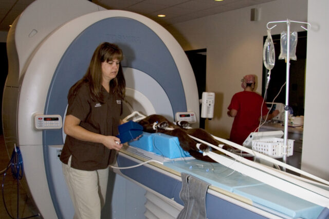
Veterinary radiology technician Donna Graber positions an animal for MRI at UF’s Veterinary Medical Center (Photo by Mark Hoffenberg)
Private and specialty practice veterinarians will soon have direct access to the Southeast's most advanced imaging diagnostics at the University of Florida Veterinary Medical Center, without needing to refer cases through the center's traditional clinical services.
The new outpatient imaging service, known as GatorVetImaging, will begin July14 and will allow veterinarians in Florida and throughout the Southeast the ability to take advantage of the same state-of-the-art imaging technologies used by UF veterinary faculty, specifically magnetic resonance imaging and CT.
"GatorVetImaging brings the best medical technology of the VMC directly to practitioners," said Matthew Winter, D.V.M., a board-certified veterinary radiologist who heads the VMC's radiology service. "We envision this as a way to assist the veterinary community in handling their more challenging and involved cases within the context of their established client/patient relationships."
While many veterinarians will continue to rely on UF as a traditional referral center offering complete patient workup, the new imaging service streamlines the diagnostic process for those veterinarians who desire only the advanced imaging piece of the patient care package at UF and wish to maintain direct primary care responsibility for their patients.
"We truly believe the new outpatient imaging service will meaningfully advance the veterinary profession," said Jim Thompson, D.V.M., Ph.D., the UF College of Veterinary Medicine's executive associate dean. "We're talking about a client-oriented service that is both efficient and cost-effective. It's a win-win for practitioners and for us at UF because we are fortunate enough to have this technology housed at our facility."
The VMC's 1.5 Tesla Toshiba Titan MR unit and the 8-slice Toshiba Acquilion CT unit at UF are the most powerful imaging tools currently available for veterinary diagnostics in the southeastern United
States. Both capabilities allow for rapid imaging with exceptional contrast and spatial resolution.
The MR unit allows highly detailed images to be obtained in multiple planes of bone and soft tissue in all species. Foot, fetlock, suspensory joints, carpus, hock and heads are regions capable of being examined through MR in the horse, while spiral CT may be used for 3-dimensional reconstruction in fracture repair planning. In small animals, both modalities are routinely applied to neurologic and orthopedic cases at the VMC, with additional studies performed for radiation planning and metastasis evaluations.
"MR allows for exquisite distinction between normal and abnormal tissues," Winter said. "The use of specialized sequences further increases the ability to distinguish between different types of pathology, ranging from hemorrhagic infarctions to primary brain tumors and inflammatory disorders."
Winter added that MR also reveals bone, tendon and ligament pathology and can show bruising, meniscal damage and ligament tears that go undetected when using traditional radiography.
"All of our radiologists have strong interests in cross-sectional imaging, which gives UF a unique ability to serve the advanced imaging needs of Florida veterinarians." Winter said.
To schedule cases, veterinarians will need to contact the GatorVetImaging coordinator to arrange an appointment. Pre-anesthesia and imaging request forms can be faxed from UF to the scheduling veterinarian, or may be downloaded from the GatorVetImaging Web site at www.GatorVetImaging.com.
Horse owners will be asked to bring their animals to UF the day prior to the scheduled procedure, which will take place the following morning. Small animals will be able to be imaged and discharged on the same day.
At the time of discharge, the animal's owner will receive a folder with a CD containing the images, as well as printed photos showing some of the more significant images from the scan. The owner will meet again with the point-of-contact clinician, who will provide instructions to follow up with the attending veterinarian regarding the next step in patient care.
A VMC radiologist will interpret the images within 48 hours of the imaging procedure, and will fax or e-mail a PDF of the results to the veterinarian. A copy of the results, and a CD with all the images, will be mailed as well.
For more information about GatorVetImaging, go to www.GatorVetImaging.com.
About the author
