Mammogram - calcifications
Definition
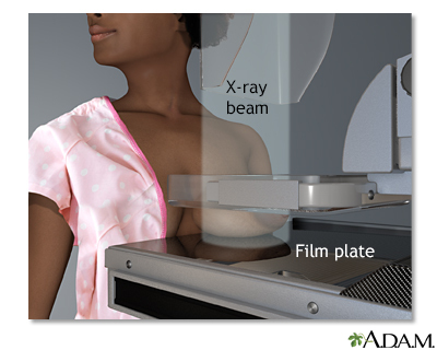
Calcifications are tiny deposits of calcium in your breast tissue. They are often seen on a mammogram.
Alternative Names
Microcalcifications or macrocalcifications; Breast cancer - calcifications; Mammography - calcifications
Information
The calcium you eat or take in as medicine does not cause calcifications in the breast.
Most calcifications are not a sign of cancer. Causes may include:
- Calcium deposits in the arteries inside your breasts
- History of breast infection
- Noncancerous (benign) breast lumps or cysts
- Past injury to the breast tissue
- Fat necrosis (breast tissue damage, usually from injury or trauma)
Large, rounded calcifications (macrocalcifications) are common in women over age 50. They look like small white dots on the mammogram. They are most likely not related to cancer. You will rarely need more testing.
Microcalcifications are tiny calcium specks seen on a mammogram. Most of the time, they are not cancer. However, these areas may need to be checked more closely if they have a certain appearance on the mammogram.
WHEN IS FURTHER TESTING NEEDED?
When microcalcifications are present on a mammogram, the doctor (a radiologist) may ask for a larger view so the areas can be examined more closely.
Calcifications that do not appear to be a problem are called benign. No specific follow-up is needed. But, your health care provider may recommend that you get a mammogram each year.
In some cases, calcifications that are slightly abnormal but do not look like a problem (such as cancer) are also called benign. Most women will need to have a follow-up mammogram in 6 months.
Calcifications that are irregular in size or shape or are tightly clustered together, are called suspicious calcifications. Your provider will recommend a stereotactic core biopsy. This is a needle biopsy that uses a type of mammogram machine to help find the calcifications. The purpose of the biopsy is to find out if the calcifications are benign (not cancer) or malignant (cancer).
Most women who have suspicious calcifications do not have cancer.
Gallery
References
Ikeda DM, Miyake KK. Mammographic analysis of breast calcifications. In: Ikeda DM, Miyake KK, eds. Breast Imaging: The Requisites. 3rd ed. St Louis, MO: Elsevier; 2017:chap 3.
James JJ, Evans AJ. The breast. In: Adam A, Dixon AK, Gillard JH, Schaefer-Prokop CM, eds. Grainger & Allison's Diagnostic Radiology. 7th ed. Philadelphia, PA: Elsevier; 2021:chap 63.
Siu AL; US Preventive Services Task Force. Screening for breast cancer: US Preventive Services Task Force recommendation statement. Ann Intern Med. 2016;164(4):279-296. PMID: 26757170 pubmed.ncbi.nlm.nih.gov/26757170/.
