Update Location

My Location

Update your location to show providers, locations, and services closest to you.

Enter a zip code
Or
Select a campus/region

Coal worker's pneumoconiosis

Definition

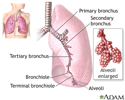
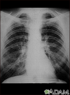
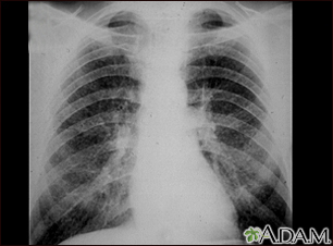
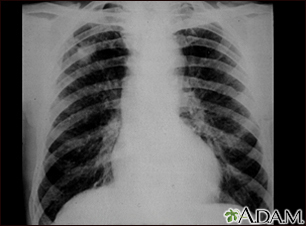
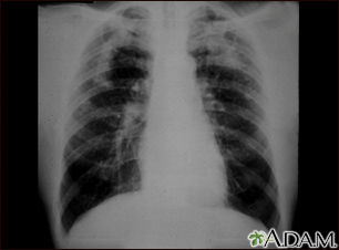
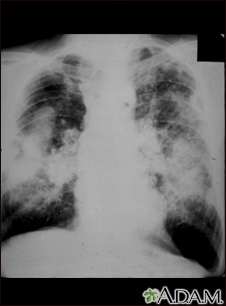
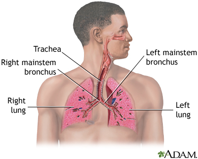
Coal worker's pneumoconiosis (CWP) is a lung disease that results from breathing in dust from coal, graphite, or man-made carbon over a long time.

CWP is also known as black lung disease.

Alternative Names

Black lung disease; Pneumoconiosis; Anthrosilicosis

Causes

CWP occurs in two forms: simple and complicated (also called progressive massive fibrosis, or PMF).

Your risk for developing CWP depends on how long you have been around coal dust. Most people with this disease are older than 50. Smoking does not increase your risk for developing this disease, but it may have an added harmful effect on the lungs.

If CWP occurs with rheumatoid arthritis, it is called Caplan syndrome.

Symptoms

Symptoms of CWP include:

Exams and Tests

The health care provider will perform a physical examination and ask about your symptoms.

Tests that may be done include:

Treatment

Treatment may include any of the following, depending on how severe your symptoms are:

  • Medicines to keep the airways open and reduce mucus
  • Pulmonary rehabilitation to help you learn ways to breathe better
  • Oxygen therapy
You should also avoid further exposure to coal dust.

Support Groups

Ask your provider about treating and managing coal worker’s pneumoconiosis. Information can be found at the American Lung Association: Treating and Managing Coal Worker's Pneumoconiosis website: www.lung.org/lung-health-diseases/lung-disease-lookup/black-lung/treating-and-managing

Outlook (Prognosis)

Outcome for the simple form is usually good. It rarely causes disability or death. The complicated form may cause shortness of breath that worsens over time.

Possible Complications

Complications may include:

When to Contact a Medical Professional

Call your provider right away if you develop a cough, shortness of breath, fever, or other signs of a lung infection, especially if you think you have the flu. Since your lungs are already damaged, it's very important to have the infection treated right away. This will prevent breathing problems from becoming severe, as well as further damage to your lungs.

Prevention

Wear a protective mask when working around coal, graphite, or man-made carbon. Follow directions to prevent high-level exposure. Companies should enforce the maximum permitted dust levels. Avoid smoking.

Gallery

Lungs
The major features of the lungs include the bronchi, the bronchioles and the alveoli. The alveoli are the microscopic blood vessel-lined sacks in which oxygen and carbon dioxide gas are exchanged.
Aortic rupture - chest X-ray
Aortic rupture (a tear in the aorta, which is the major artery coming from the heart) can be seen on a chest X-ray. In this case, it was caused by a traumatic perforation of the thoracic aorta. This is how the X-ray appears when the chest is full of blood (right-sided hemothorax) seen here as cloudiness on the left side of the picture.
Coronavirus
Coronaviruses are a family of viruses. Infection with these viruses can cause mild to moderate respiratory illnesses such as the common cold. Some coronaviruses may cause severe illness and lead to pneumonia or even death.

References

Go LHT, Cohen RA. Pneumoconioses. In: Broaddus VC, King TE, Ernst JD, et al, eds. Murray and Nadel's Textbook of Respiratory Medicine. 7th ed. Philadelphia, PA: Elsevier; 2022:chap 101.

Tarlo SM. Occupational lung disease. In: Goldman L, Schafer AI, eds. Goldman-Cecil Medicine. 26th ed. Philadelphia, PA: Elsevier; 2020:chap 87.

Last reviewed May 30, 2021 by Denis Hadjiliadis, MD, MHS, Paul F. Harron, Jr. Associate Professor of Medicine, Pulmonary, Allergy, and Critical Care, Perelman School of Medicine, University of Pennsylvania, Philadelphia, PA. Also reviewed by David Zieve, MD, MHA, Medical Director, Brenda Conaway, Editorial Director, and the A.D.A.M. Editorial team..

Related specialties

Aftercare and more