Cloudy cornea
Definition
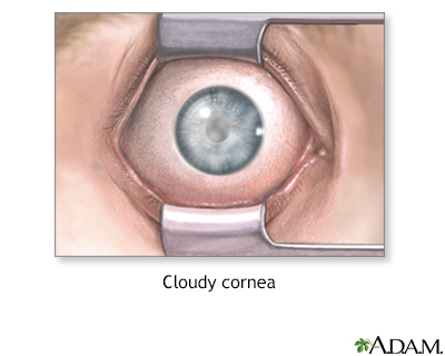
A cloudy cornea is a loss of transparency of the cornea.
Alternative Names
Corneal opacification; Corneal scarring; Corneal edema
Causes
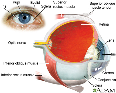
The cornea is the front wall of the eye. It is normally clear. It helps focus the light entering the eye.
Causes of cloudy cornea include:
- Inflammation
- Sensitivity to non-infectious bacteria or toxins
- Infection
- Keratitis
- Trachoma
- River blindness
- Corneal ulcers
- Swelling (edema)
- Acute glaucoma
- Birth injury
- Fuchs dystrophy
- Dryness of the eye due to Sjögren syndrome, vitamin A deficiency, or LASIK eye surgery
- Corneal dystrophy (inherited metabolic disease)
- Keratoconus
- Injury to the eye, including chemical burns and welding injury
- Tumors or growths on the eye
- Pterygium
- Bowen disease
Clouding may affect all or part of the cornea. It leads to different amounts of vision loss. You may not have any symptoms in the early stages.
Home Care
Contact your health care provider. There is no appropriate home care.
When to Contact a Medical Professional
Contact your provider if:
- The outer surface of your eye appears cloudy.
- You have trouble with your vision.
Note: You will need to see an ophthalmologist for vision or eye problems. However, your primary care provider may also be involved if the problem could be due to a whole-body (systemic) disease.
What to Expect at Your Office Visit
The provider will examine your eyes and ask about your medical history. The two main questions will be if your vision is affected and if you have seen a spot on the front of your eye.
Other questions may include:
- When did you first notice this?
- Does it affect both eyes?
- Do you have trouble with your vision?
- Is it constant or intermittent?
- Do you wear contact lenses?
- Is there any history of injury to the eye?
- Has there been any discomfort? If so, is there anything that helps?
Tests may include:
- Biopsy of lid tissue
- Computer mapping of the cornea (corneal topography)
- Schirmer's test for eye dryness
- Special photographs to measure the cells of the cornea
- Standard eye exam
- Ultrasound to measure corneal thickness
Gallery
References
Cioffi GA, Liebmann JM. Diseases of the visual system. In: Goldman L, Schafer AI, eds. Goldman-Cecil Medicine. 26th ed. Philadelphia, PA: Elsevier; 2020:chap 395.
Goncalves De Pinho AR. Corneal tissue engineering: new applications for corneal stromal stem cells (Doctoral dissertation, UCL (University College London)). discovery.ucl.ac.uk/id/eprint/10127500/1/PhD_FinalThesis_AnaRitaPinho.pdf. Published December 2020. Accessed November 4, 2022.
Guluma K, Lee JE. Ophthalmology. In: Walls RM, ed. Rosen's Emergency Medicine: Concepts and Clinical Practice. 10th ed. Philadelphia, PA: Elsevier; 2023:chap 57.
Kane JS, Kane SA. Cloudy corneas, plus. J Pediatr Ophthalmol Strabismus. 2022;59(2):73. PMID: 35343823 pubmed.ncbi.nlm.nih.gov/35343823/.
Kataguiri P, Kenyon KR, Batta P, Wadia HP, Sugar J. Corneal and external eye manifestations of systemic disease. In: Yanoff M, Duker JS, eds. Ophthalmology. 5th ed. Philadelphia, PA: Elsevier; 2019:chap 4.25.
Lisch W, Weiss JS. Early and late clinical landmarks of corneal dystrophies. Exp Eye Res. 2020;198:108139. PMID: 32726603 pubmed.ncbi.nlm.nih.gov/32726603/.
Patel SS, Goldstein DA. Episcleritis and scleritis. In: Yanoff M, Duker JS, eds. Ophthalmology. 5th ed. Philadelphia, PA: Elsevier; 2019:chap 4.11.
