Bone x-ray
Definition
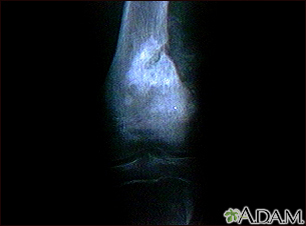
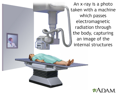
A bone x-ray is an imaging test to look at the bones.
Alternative Names
X-ray - bone
How the Test is Performed
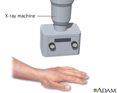
The test is done in a hospital radiology department or in the health care provider's office by an x-ray technician. For the test, you will position the bone to be x-rayed on the table. Pictures are then taken, and the bone is repositioned for different views.
How to Prepare for the Test
Tell the health care provider if you are pregnant. You must remove all jewelry for the x-ray.
How the Test will Feel
The x-rays are painless. Changing position for getting different views of the bone may be uncomfortable.
Why the Test is Performed
A bone x-ray is used to look for injuries or conditions affecting the bone.
What Abnormal Results Mean
Abnormal findings include:
- Fractures or broken bone
- Bone tumors
- Degenerative bone conditions
- Osteomyelitis (inflammation of the bone caused by an infection)
Additional conditions under which the test may be performed:
Risks
There is low radiation exposure. X-ray machines are set to provide the smallest amount of radiation exposure needed to produce the image. Most experts feel that the risk is low compared with the benefits.
Children and the fetuses of pregnant women are more sensitive to the risks of the x-ray. A protective shield may be worn over areas not being scanned.
Gallery
References
Kapoor G, Toms AP. Current status of imaging of the musculoskeletal system. In: Adam A, Dixon AK, Gillard JH, Schaefer-Prokop CM, eds. Grainger & Allison's Diagnostic Radiology: A Textbook of Medical Imaging. 7th ed. Philadelphia, PA: Elsevier; 2021:chap 38.
Contreras F, Perez J, Jose J. Imaging overview. In: Miller MD, Thompson SR. eds. DeLee, Drez, & Miller's Orthopaedic Sports Medicine. 5th ed. Philadelphia, PA: Elsevier; 2020:chap 7.