Like A Water Balloon in a River
When Madeline Lee went to her 28-week prenatal checkup, she was not able to see her son’s head in the sonograms. Her doctor later determined that the fetus had…

Update your location to show providers, locations, and services closest to you.
The most common type of brain cyst, arachnoid cysts are usually congenital, or present at birth. These cysts are called primary arachnoid cysts. Arachnoid cysts that develop later in life are called secondary arachnoid cysts. These are not brain tumors, but are rather benign sacs usually filled with clear cerebrospinal fluid (CSF).
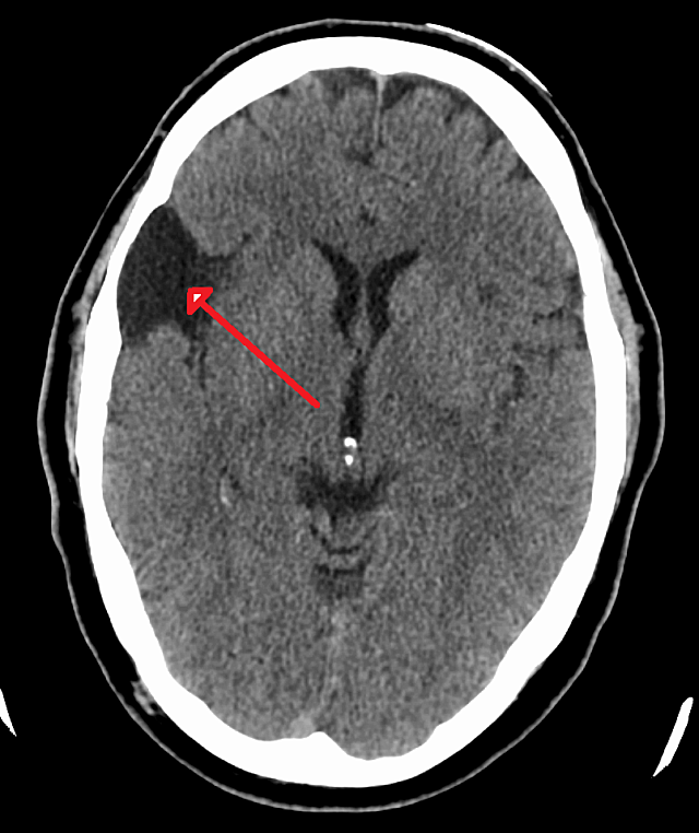
There are three layers of tissue that surround the brain and spinal cord. The middle layer is called the arachnoid membrane, and this is where arachnoid cysts develop. Arachnoid cysts usually occur in the brain, but arachnoid cysts can appear in the spinal cord as well.
The walls of an arachnoid cyst block CSF from draining naturally into your brain, causing the fluid to remain inside the sac. If they continue to retain this fluid, they have the potential to grow to a large size and put pressure on the brain and lead to several issues.
There are many ways to treat arachnoid cysts, from simply watching them over time to placement of a drainage shunt tube, and various types of surgery to eliminate the cyst or prevent its growth. UF’s neurosurgeons have specialized training in these operations and can even offer the latest in minimally invasive arachnoid cyst surgery using tiny cameras and other technical advancements.
Arachnoid cysts are typically asymptomatic, but they do present symptoms in some cases. These signs depend on the location and size of the cyst.
Those located in the brain may produce one or more of the following symptoms:
Those located in the spinal column may cause the following symptoms:
Arachnoid cysts can also bring on other conditions. Hydrocephalus, an accumulation of excessive cerebrospinal fluid in the brain, can produce increased cranial pressure. Macrocephaly, an abnormally enlarged head, can occur in rare cases due to a malformation of certain cranial bones, especially in children.
Neurological symptoms can also appear, such as developmental delays, behavioral changes, inability to control voluntary movements (ataxia), difficulties with balance and walking, cognitive impairment, and weakness or paralysis on one side of the body (hemiparesis).
The exact cause of a primary, or congenital, arachnoid cyst is unknown. It arises from an abnormal growth of the arachnoid membrane in a fetus during pregnancy. There’s a possibility they may be genetic, although they rarely run in families.
Secondary arachnoid cysts, also known as noncongenital arachnoid cysts, can be caused by a few things:
Diagnosis of an arachnoid cyst can come from imaging tests such as a CT or MRI scan, which help doctors pinpoint the location and traits of the cyst. Since most arachnoid cysts do not cause symptoms, they are also frequently discovered by accident when a person undergoes a brain scan for an unrelated reason (i.e., after a car crash or a stroke).
Some arachnoid cysts, even large ones, may not cause symptoms or put pressure on the brain or spinal cord. In those cases, your doctor may advise you to leave it untreated. Instead, the cyst will likely be monitored over time to watch for any growth or other changes. If problems develop, treatment options will be explored.
Treating an arachnoid cyst mainly revolves around draining fluid from the cyst and relieving pressure, and this can be achieved through a number of treatment options. If the cyst resides in the brain, your doctor will likely drain it. In this case, one of two procedures can be performed:
Symptomatic cysts in the spinal column may be removed entirely with surgery. If not, one of the two aforementioned procedures can be applied to drain it.
Those with an asymptomatic cyst will likely enjoy a normal life, and may not even require treatment. Your doctor may only require regular check-ups to observe the cyst for changes.
Those with a symptomatic cyst can be treated by draining or removing it completely, which should address any symptoms. There are rare cases in which untreated, expanding arachnoid cysts can lead to permanent neurological damage as it puts pressure on surrounding brain tissue.

When Madeline Lee went to her 28-week prenatal checkup, she was not able to see her son’s head in the sonograms. Her doctor later determined that the fetus had…